


 Back
Back


Computed tomography (CT) is often associated with magnetic resonance imaging (MRI). However, there are still differences between these research methods. These differences will be covered in this article. Just like on x-rays, bones and structures that contain calcium appear white on computed tomography; soft tissues (e.g. heart) - in shades of gray. Tissues that are close in density to air (such as lungs, intestines) are displayed in black. Thanks to state-of-the-art computer technology, a computed tomography session is carried out so quickly that even infants and young children do not need to be anesthetized or injected with sedatives to lie quietly.
In some cases, a contrast agent is injected intravenously to diagnose a tumor, for example, to distinguish a tumor from a postoperative scar. It is administered either through a central venous catheter, which is absolutely painless.
What is CT?
Computed tomography (CT) is a special technique that uses X-rays. With its help, the human body is translucent with X-rays. In children and adolescents with cancer, doctors use computed tomography for preliminary diagnosis and to monitor treatment results, as well as when planning surgery and radiation therapy (radiation). The study is prescribed independently or in addition to MRI (magnetic resonance imaging). It is also used for emergency diagnostics. The results of computed tomography can be obtained fairly quickly.
What is the difference between CT and MRI?
Computed tomography is the best way to perceive bone structures. It is for this reason that CT is more commonly used to identify bone injuries and various injuries.
On MRI, soft tissues are clearly visible: muscles, blood vessels, cartilage, spinal cord and brain. Therefore, MRI is best used to detect tumors and pathologies in the field of neurology and neurosurgery.
There are also differences in the way these procedures operate.
Under the influence of X-rays on a computed tomography (CT) scanner, a series of images of the object under study is obtained.
A magnetic resonance imaging (MRI) scanner operates because of a magnetic field in which a patient is placed.
MRI can help you get results that are more accurate in the following cases:
- The body's response to the injected contrast agent was detected during CT
- You should check the condition of the brain, the condition of soft tissues
- Musculoskeletal diseases of children
- It is necessary to check the condition of the pituitary gland, the condition of the nerve cells of the brain
- Suspected cancer
Preparation to CT
Not all types of computed tomography require special preliminary preparation.
Preparation for computed tomography of the pelvic organs: one day before the study, exclude from the diet foods that contribute to increased gas production (black bread, fermented milk, food containing coarse fibers, and so on). On the day before the study, cleanse the intestines: at home, it is advisable to do a cleansing enema; for women, before conducting a study, a cotton swab (not tight) is inserted into the vagina - dry or moistened with a contrast agent; the study begins with a full bladder.
Preparation for computed tomography of the abdominal cavity and retroperitoneal space: one day before the study: exclude from the diet foods that contribute to increased gas production (black bread, fermented milk, food with coarse fibers, and so on). On the eve of the study in the evening, a light dinner is allowed: liquid food; the study is carried out on an empty stomach: in the morning on the day of the study, it is necessary to exclude food intake (do not have breakfast). If it is allowed to drink tea for more than 2 hours before the study; when conducting a study in the afternoon, the last light meal should be at least 5 hours in advance (fasting interval of 5 hours or more); for research, have 1 liter of still drinking water with you.
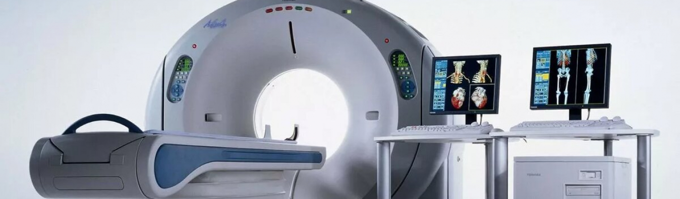
How does the CT work?
During the examination, the patient lies motionless. The X-ray beam is directed by the computer and rotates around it, shining through the body from different directions. X-ray images are taken from different angles, they form a single picture. In a matter of seconds, the computer processes information from thousands of images. The images in these images are taken from various cross-sections of the body, in centimeter or millimeter increments. Then, a high-precision detailed image is printed. For some situations, for example, when a complex operation is planned, the information obtained is converted into a three-dimensional image (3D).
CT advantages
CT method guarantees clearer visualization of bone structures, vessel walls, acute intracranial bleeding, ultrastructure of the lungs. In addition, CT medical examinations have shorter exam times than magnetic resonance imaging. CT is also an optimal alternative for patients for whom an MRI examination is contraindicated, for example, if the patient has pacemakers and metal implants in the patient's body, as well as if there is a fear of confined spaces. It is CT that provides improved tissue and organ differentiation through high-resolution cross-sectional images without layering.
Contraindications to CT
Computed tomography is painless, but it has radiation exposure (dose of radiation). The radiologist always analyzes the results together with the attending physicians. Except for life-threatening emergencies, computed tomography for children and adolescents under the age of 18 may only be performed with the written permission of their parents. According to German law, the responsible radiologist is obliged to inform the parents of the possible risks beforehand.
What diseases can CT reveal?
The effectiveness of computed tomography has been proven in the study of such systems and organs as:
- lungs, bronchi;
- brain;
- organs of the small pelvis;
- spine and other parts of the skeleton;
- vessels;
- skin, subcutaneous tissue;
- abdominal organs;
- lymph nodes.
Accordingly, diseases of these organs will be visible on computed tomography.
Types of CT
CT of the temporal bones is performed to diagnose the pathology of this anatomical formation with hearing loss, dizziness, and pathology of the organ of balance. Does not require (with rare exceptions) the introduction of contrast agents. Non-alternative method of radiation diagnostics. Spiral CT is preferred.
There are two types of CT of the brain.
- without amplification, that is, without intravenous administration of a radiopaque contrast agent. It is carried out with suspicion of a pathology developing in the brain and its membranes. It is used mainly as a screening method to search for probable pathology. With a non-neoplastic nature of the pathological changes found with it, as a rule, it does not require the use of other computed tomography techniques.
- with intravenous enhancement – performed if a brain tumor, vascular malformations and aneurysms are suspected. Intravenous injection of a radiopaque (CT image contrast enhancement technique) substance can be performed 3-5 minutes before the start of the CT scan or after native CT. In case of damage to the blood-brain barrier and / or the presence of a pathological vascular network due to an increase in the X-ray density of the pathological focus, it is possible to recognize the presence and localization of the pathological process. Does not allow you to see the feeding vessel. Additional computed tomography technique.
Orbital CT is done to assess the condition of the bony walls and contents. The information (including the 3D picture) about the bones that make up the orbit is unique. CT allows visualizing the eyeball, optic nerve, rectus muscles of the eye, and adipose tissue. Highly informative for orbital pseudotumors. Together with ultrasound diagnostics, it is the method of choice for eye and orbit injuries. May be the main method of radiation diagnostics. Spiral CT is preferred.
CT of the chest (spiral) allows you to obtain images of the ribs, sternum, vertebrae and register the violation of their activity in developmental anomalies and injuries in conditions of three-dimensional modeling (3D reconstruction). It is especially indicated for chest injuries, combined chest and abdominal injuries.
CT of the pelvic organs is used to search for and differential diagnosis of pathological changes in the parenchymal pelvic organs. Unique in terms of assessing anatomical relationships. Clarifying method of radiation diagnostics. It is somewhat inferior in informational value of echoscopy in pathology of the bladder, prostate, ovaries, uterus. It is most informative for recognizing the pathology of the soft tissues of the small pelvis, and when using spiral CT with contrast, for detecting enlarged lymph nodes. Clarifying method of radiation diagnostics. Spiral CT is preferred.
A CT of the spine and segments is performed to assess the condition of the vertebrae and intervertebral discs. The information content of CT in osteochondrosis, spondylosis, other pathology of the bodies and processes of the vertebrae, intervertebral discs, ligaments of the spinal column is slightly higher than MRI. MRI does not compete in visualization of pathological changes in the spinal cord. Method of choice for radiological diagnosis of osteochondrosis, vertebral fractures. Spiral CT is preferable because all parts of the spine can be scanned in one examination.
CT of joints and bones is performed to detect pathological processes in the bones that make up the joint, its soft tissues and, in the case of the knee joint, its ligaments. In some cases, the information obtained with CT is unique. It is inferior to MRI in visualizing cartilage and ligaments, but superior in visualizing bone tissue (with MRI, bone trabeculae are not visible at all). Clarifying diagnostic method. Preferred helical CT in thin slices.
CT of the neck is performed for the diagnosis and differential diagnosis of neoplasms in this area, for determining the degree of tumor invasion of large vessels (the main method of radiological diagnosis), for assessing the state of the lymphatic catch of the neck (for informational content it is equal to echoscopy).
CT of parenchymal organs of the abdomen - search and differential diagnosis of pathology of the pancreas and liver. It can be the leading (abdominal trauma), main (pathology of organs and tissues of the retroperitoneal space) or additional (pathology of the liver, biliary tract, spleen) by the method of radiation diagnostics. It is of little use for pathology of the stomach and intestines. Spiral CT of the abdomen may be the first and preferred method of radiological diagnosis of the state of its organs and the method of choice in patients in severe condition and with trauma, including combined. The most effective CT scan is with the introduction of a contrast agent using an automatic syringe.
Medical centers offering CT services in Belarus:
- LODE
- State Institution Republican Clinical Medical Center of the Administrative Department of the President of the Republic of Belarus
- Medical center “Kravira” in Minsk
- Medical center “EXANA” in Minsk
- Medical center MTZ in Minsk
- Medical Center AVICENNA
- Clinic Aqua-Minsk
- Medical Center “Doctor Profi”