Where can you do an ultrasound in Minsk?
Ultrasound diagnostics is one of the leading examination methods.
If you have recently arrived in Belarus and do not know where to do an ultrasound in Minsk, the information on our website will greatly facilitate your searches, since we have collected all the best centers in the field of ultrasound diagnostics for you.
You can see the list of centers on the map, and familiarize yourself with the services provided on the link.
What is an ultrasound scan?
Ultrasound diagnostics is a method of radiation diagnostics, which uses high-frequency (ultra) sound waves to obtain images of internal organs.
The method is based on the reflection of ultrasonic waves from the internal structures of the body with subsequent registration and creating of image using a special device.
Advantages of the method
Nowadays, in all branches of medicine, the method of ultrasound diagnosis is used. It allows you to identify diseases in the early stages. The main advantages:
- Safety;
- Simplicity;
- Lack of contraindications;
- Lack of radiation exposure (therefore, ultrasound is actively used in case of pregnancy);
- Non-invasiveness (study without violation of the skin);
- High diagnostic efficiency (accuracy over 80%);
- The possibility of multiple studies;
- Conducting research in real time;
- Transportability of ultrasonic equipment;
- Study of several organs and systems at once.

Cranial ultrasound
Ultrasound of the brain structures cannot be performed in an adult, since the bones of the skull interfere with the passage of the ultrasound wave.
However, the study of cerebral vessels seems possible. Thanks to this procedure, the blood in the vessels can be examined. Basically, it is carried out together with a scan of the vessels of the neck.
The day before an ultrasound, it is recommended to refuse to take coffee.
The method allows to identify and evaluate:
- Damage to the vascular wall;
- The condition of the vascular wall;
- The presence of neoplasms in the vessels;
- Vascular elasticity.
Cardiac Ultrasound
In the early stages of the disease, the cardiovascular system is asymptomatic, the ultrasound method allows them to be detected.
The main structures that can be monitored using ultrasound:
- Cardiac chambers;
- Heart valves;
- Atrial walls;
- Walls of ventricles;
- Vascular integrity;
- Heart muscles;
- Heart bag.
The main indications for cardiac ultrasound:
- Myocardial infarction;
- Cardiac angina;
- Heart murmurs;
- Shortness of breath;
- Thrombosis;
- Heart disease.
Pelvic ultrasound
Pelvic ultrasound is a comprehensive study of the organs of the genitourinary and reproductive systems, evaluating its functional state.
There are four methods for performing pelvic ultrasound:
- Transabdominal (examination of internal organs through the abdominal wall), the study is carried out with a full bladder;
- Transvaginal (examination of internal organs by introducing a special sensor into the vagina),
- Combined. A transabdominal ultrasound is performed, followed by emptying of the bladder and the transition to the transvaginal method.
- Transrectal (examination of internal organs using a sensor that is inserted into the rectum). The method is used for girls who do not live sexually.
Using pelvic ultrasound, you can identify:
- Benign and malignant tumors in the uterus, fallopian tubes, ovaries;
- Ectopic pregnancy;
- Parameters of the dominant follicle in ovulation;
- Fetal development during pregnancy;
- The structure of the prostate gland, testicles;
- Problems during urinating.
A timely visit to a specialist and making diagnostics is the key to your health!
Ultrasound of the kidneys
Ultrasound of the kidneys is a comprehensive study of the genitourinary system, which allows you to evaluate its condition, linear blood flow velocity, detect benign and malignant neoplasms, the state of the vessels of the kidneys, as well as the structure of the organ itself.
Special preparation before the study is not required, it is carried out in the lateral position quickly and painlessly, the results are interpreted in real time.
The indications for ultrasound of the kidneys:
- Low back pain;
- Chronic and acute diseases of the bladder;
- Swelling of the body;
- Difficult and painful urination;
- Rise in blood pressure, accompanied by headaches;
- Changes in urine colour;
- Changes in the amount of urine.
As a result of an ultrasound of the kidneys, the following are revealed:
- Dimensions of the kidneys;
- The course of the inflammatory process;
- Stones, cysts;
- Benign and malignant diseases, etc.
A liver ultrasound
A liver ultrasound is a method of non-invasive study of the structure of the liver and its changes in the course of diseases of various origins.
Ultrasound examination of the liver is often carried out together with ultrasound of the abdominal cavity and elastometry - this is an ultrasound method for examining the stiffness of liver tissue.
Before the study, you must refrain from eating for 6-8 hours, since it is carried out on an empty stomach.
Indications for a liver ultrasound:
- Enlarged liver in size;
- Alcohol addiction;
- Injuries in the abdominal cavity;
- An icteric shade of eyes and skin;
- Exceeding the norms of liver enzymes, etc.
As a result of liver ultrasound, you can evaluate:
- Линейную скорость кровотока.
- The size of the lobes of the liver;
- Condition of tissues, liver structure, presence of neoplasms and seals;
- The state of the vascular system of the liver;
- Linear blood flow velocity.
Abdominal ultrasound
Ultrasound of the abdominal cavity is a comprehensive non-invasive examination of the liver, gall bladder (with and without its load), pancreas, spleen, retroperitoneal space, which allows to detect dystrophic changes in the tissues of these organs and foci of inflammation, pathology of the vascular network, stones in the gall bladder and its ducts, diseases at an early stage.
The study takes 15-20 minutes, is safe and very accurate.
Before the study, it is necessary to abstain from food for 6-8 hours, since it is carried out on an empty stomach. If you suffer from constipation, then the day before you need to make an enema; flatulence - to take 2-3 capsules of simethicone 3 times a day.
Indications for abdominal ultrasound:
- Stomach ache;
- Low back pain;
- Increased gases;
- Nausea;
- Feeling of heaviness in the stomach;
- A feeling of bitterness in the mouth;
- Enlarged liver, spleen;
- The presence of pathological formations.
As a result, of abdominal ultrasound, you can identify:
- Gallstone disease;
- Cholecystitis, pancreatitis and other inflammatory diseases of the gastrointestinal tract;
- Cirrhosis of the liver;
- Diseases at an early stage, in which there is no clinical picture;
- Gallbladder dyskinesia;
- Polyps;
- Neoplasms;
- Change in the shape and size of the abdominal organs.
Ultrasound of the stomach
Ultrasound of the stomach is a painless procedure, but not the most effective research method. Ultrasound of hollow organs, including the stomach, is difficult, because the waves of ultrasound are completely reflected from the air that is contained in it. Thus, it is impossible to assess the condition of the mucosa, the most informative method in this case is endoscopy.
The study is carried out in two stages:
- First, the doctor conducts an ultrasound of the stomach on an empty stomach, assesses the condition of the walls, the presence of contents, conducts a search for the most painful area;
- Next, the patient needs to drink half a liter of water 25-35 degrees Celsius. A doctor examines a full stomach.
Indications for ultrasound of the stomach:
- Categorical patient refusal from endoscopic examination (swallowing the probe);
- Contraindications to endoscopy;
- Pain and heaviness in the stomach;
- Change in appetite;
- Nausea, vomiting, bitterness in the mouth;
- Gastritis in acute and chronic form;
- Stomach ulcer;
- Neoplasms in the stomach;
- Monitoring of treatment.
As a result of ultrasound of the stomach, you can identify:
- Features of the location of the stomach;
- The presence of contents in the stomach on an empty stomach;
- The thickness of the gastric wall;
- Peristalsis of the stomach after filling with liquid;
- The presence of GERD (gastroesophagic reflux);
- The presence of neoplasms in the wall of the stomach, which grow in its lumen;
- The condition of the lymph nodes.
Bowel ultrasound
Ultrasound of the intestine is a safe and painless procedure, but not the most informative research method. Ultrasound of hollow organs, including the intestines, is difficult, because ultrasound waves are completely reflected from the air that is contained in it. In order to analyze the condition of the mucus, in this case, it is better to undergo a colonoscopy.
Indications for a bowel ultrasound:
- Prophylactic examination for colon cancer;
- Constipation and loose stools of a chronic nature;
- Anemia;
- Control after surgery on the stomach, gall bladder, to remove appendicitis;
- Suspicion of appendicitis;
- Enterocolitis, pseudomembranous colitis and other inflammatory bowel diseases;
- Mesenteric lymphadenitis.
As a result of an intestinal ultrasound, you can identify:
- Pathology in tissues located near the intestine;
- The most painful area (by palpation);
- The thickness of the intestinal wall;
- Changes in the intestinal wall;
- The presence of free fluid in the abdominal cavity;
- Enlarged lymph nodes.
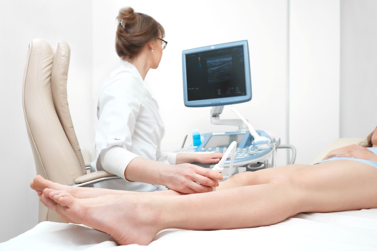
Ultrasound of the legs
Throughout life, a person has to move a lot, recently the pace of life has been increasing more and more. The joints are under significant stress. With age, people experience pain in the joints of the legs. Most of them suffer from ankles. The negative results of excessive load are edema, inflammation, dislocation.
With an unknown etiology of ankle damage, ultrasound is the most informative method.
When performing an ultrasound of the legs, the doctor can determine the cause of the inflammation, as well as the degree of injury.

Ultrasound of the joints
At the moment, ultrasound of the joints is prescribed in combination with elastography. Elastography is a method in which an ultrasound beam is emitted by a special apparatus, reflected from tissues and recorded on the device.
The essence of the method is that healthy tissues and tissues with tumours have different elasticities (the latter are more dense). This combination of methods allows you to assess the condition:
- Cartilage;
- Ligaments;
- Tendons;
- Joint bags;
- Production of joint fluid.
Indications for ultrasound of the joints are:
- Pain and mobility limitations;
- Injuries and inflammatory processes in the joints;
- Pathologies of an endocrine nature;
- Monitoring the effectiveness of therapy for joint diseases.
There is no need for preparation for this procedure.
Ultrasound of vessels, veins and arteries of the lower extremities
The vascular system is extremely important for the human body, because through this extensive network, important substances are delivered to each cell of the body. However, sometimes this transport mechanism fails. To understand the possible reasons allows the method of ultrasound of blood vessels, veins and arteries of the lower extremities.
During the study, the doctor assesses the condition of the walls of the vessels, their patency, the presence / absence of blood clots, the condition of the surrounding tissues.
Indications for ultrasound of blood vessels, veins and arteries of the lower extremities are:
- Thrombophlebitis;
- Varicose veins;
- Numbness of the lower extremities;
- Heaviness and swelling of the legs;
- Pain and cold feet;
- Diabetes;
- Chronic venous insufficiency.
The procedure does not require special preparation, its duration is no more than 50 minutes.
Ultrasound of the prostate
Ultrasound of the prostate is an important and high-demanded study. During it, the doctor can assess the size of the organ, its structure, identify the presence of benign/malignant neoplasms, the causes of impaired potency and infertility.
There are two ways to perform an ultrasound of the prostate gland:
- Transabdominally - through the abdominal wall. The patient does not need preparation, the only condition is a full bladder. This option is not suitable for patients with urinary incontinence and is less informative.
- Transrectally - the patient lies on his side, a condom is put on a special sensor, a lubricant is applied, and it is inserted into the rectum. This method is highly informative and most preferred for diagnosis, since the sensor is adjacent directly to the organ.
The patient needs preparation before the study: abstinence from eating 6-8 hours, as well as setting an enema 1-1.5 hours before the procedure in order to remove feces from the rectum. Contraindications will be inflammatory diseases of the rectum (hemorrhoids, tumors and hemorrhoidal fissures).
Indications for ultrasound of the prostate gland:
- Pain in the groin;
- Pain in the lower abdomen;
- Inflammation of the urethra;
- Inflammation of the prostate gland;
- STIs (sexually transmitted infections);
- Frequent urination;
- The presence of blood in semen/urine;
- Infertility;
- Violation of potency.
Breast ultrasound
Due to the deterioration of the environmental situation, diet, increase in the number of stresses, the number of neoplasms is growing. Breast ultrasound is an extremely important examination for every woman. It can be carried out together with elastography (allows you to detect seals in the tissues).
The method is safe and painless, and also makes it possible to detect formations up to 5 mm, which cannot yet be determined by palpation.
It is recommended to undergo ultrasound of the mammary glands for prophylactic purposes at least 1 time per year. And for women and girls with a family history of breast cancer, at least once every 6 months.
Indications for an ultrasound of the breast:
- The presence of seals on palpation, both at the examination of the doctor, and the patient independently;
- Discharge from the nipples;
- Change in the areola of the nipple;
- Peeling of the nipples;
- Enlarged lymph nodes in the armpit;
- Monitoring the development of recurrence of neoplasm growth after surgical removal;
- Mastopathy.
Thyroid ultrasound
Ultrasound of the thyroid gland is a simple, quick and informative research method. It allows you to evaluate blood flow, both in the gland and in the surrounding lymphatic vessels. It is often carried out in combination with elastography (a method that allows you to evaluate the density of tissues for the presence of neoplasms in the thyroid gland).
The patient is placed on a couch, gel is applied to the neck and a special sensor is used to examine the position of the gland, size, structure, shape, the presence of "nodes", evaluate blood flow in the organ, as well as in formations that are more than 3-4 mm in size.
Indications for ultrasound of the thyroid gland are:
- The presence of neoplasms;
- Preventive examination (especially for people: after 40 years; taking hormonal drugs; having a predisposition to thyroid disease and diabetes).
- Nervousness, hand tremor;
- Fast fatiguability;
- Weight change;
- Hair loss;
- Difficulty swallowing (sensation of a lump in the throat).
Ultrasound of the bladder
Ultrasound of the bladder is a safe and informative research method.
There are several ways to carry it out:
- Transabdominally (through the abdominal wall);
- Transrectally (through the rectum, more often used for men in conjunction with ultrasound of the prostate gland);
- Transvaginally (used for women, often together with pelvic ultrasound);
- Intracavitary method (through the urethra for both sexes);
An ultrasound of the bladder is carried out prophylactically to control the prescribed therapy, as well as immediately in cases of:
- Injuries to the lower abdomen;
- Pain during urination;
- Change in the color of urine and the appearance of flakes in it, sediment.
When conducting an ultrasound of the bladder, you can identify:
- Anomalies in the development of the bladder;
- Urological diseases;
- Urolithiasis and the nature of stones, their number;
- Benign and malignant tumors;
- Diverticulum - pathological protrusion of the bladder wall;
- Foreign bodies;
- Other disorders of the genitourinary system.
Before the procedure, special training is required. Three days you must refrain from taking raw fruits and vegetables, bakery products, dairy products and carbonated drinks.
With increased gas formation, it is necessary to take Simethicone preparations 2 capsules three times a day the day before the procedure.
An hour and a half before the procedure, men need to drink 1-1.5 liters of water, women - up to 1 liter, pregnant women - 500 ml.
Soft tissue ultrasound
Ultrasound of the soft tissues is a comprehensive diagnosis of the skin, subcutaneous fat, ligaments, tendons, muscles and joints. The research method allows you to identify diseases in the early stages, in case of injury - to evaluate the speed of blood flow, as well as the volume of fluid.
It is safe, highly informative, affordable.
Indications for ultrasound of soft tissues:
- Seals palpation;
- Hernia;
- Hematomas;
- Purulent inflammations and abscesses;
- Monitoring the dynamics of the prescribed treatment;
- Rupture of ligaments and tendons;
- Fractures.
The study does not require special preparation.
Ultrasound during pregnancy
Ultrasound during pregnancy is an important diagnostic method that allows a healthy baby to be born.
There are several methods for ultrasound for pregnant women. They differ depending on the purpose of obtaining information:
- Sonography - allows you to assess the condition of the placental system;
- Ultrasonic fetometry - allows you to assess the development of the fetus throughout pregnancy, to diagnose growth retardation, and also makes it possible to determine the state of various organ systems (respiratory, urinary).
- Antenatal echocardiography - allows you to get information about the fetal heart and blood circulation;
- Dopplerography - allows you to assess the state of blood flow in the fetoplacental system.
Ultrasound should be done on the following dates:
- At the first sign of pregnancy;
- At 12-14 weeks. At this time, it is possible to identify an undeveloped pregnancy, an ectopic pregnancy, a threat of miscarriage, etc;
- On the 24th week. At this time, when examining the head of the fetus and ventricles of the brain, it is possible to identify disorders in the development of the central nervous system (for example, hydrocephalus);
- At 32-34th week. At this time, all organs and systems are already being formed, the condition of which is assessed by the doctor using ultrasound in combination with dopplerography.
Pediatric ultrasound
Ultrasound is performed at any age and has no contraindications.
Ultrasound examinations of children are prescribed for various indications, but pediatricians especially recommend a neurosonography procedure. This is an ultrasound of the brain, which is performed for one ear old children, until they have densely fused skull bones, in order to control the development of parts of the brain, the presence of neoplasms, defects.
In Minsk, you can do a children's ultrasound in the following centers: Eksana, Okomedson, Astra, Grandmedica and others.
How to make an appointment with a doctor for an ultrasound scan in Minsk?
You can sign up for an ultrasound procedure in a clinic at your place of residence or call one of the private clinics where you will be notified of the prices for services and the doctor’s work hours.

Medical centers of Minsk
We have compiled for you a list of medical centers in Minsk where you can go through an ultrasound procedure.
- Sedmoye nebo
Address: st. Filimonova, 53.
- Eksana
Address: st. Mogilev, 5/1.
- Kletochnye Tehnologii
Address: st. P. Brovka 15A-2.
- Cardian
Address: st. International, 10.
- Medclinic
Address: st. Pritytsky 79.
- Kravira
Address: Ave. Pobediteley, 45.
- Okomedson
Address: st. Pritytsky, 91 pom. 434.
- 1st Central Regional Clinical Polyclinic of the Central District of Minsk
Address: st. Suhaya, 6.
- Center N. N. Alexandrova
Address: Minsk region, ag. Lesnoy
- Center Sna
Address: Independence Avenue, 72a, apt. 1H.
- K-aktiv
Address: st. Ponomarenko, 35a.
- Lubimy doctor
Address: st. Matusevich, 70.
- Ultrasound medical center of Dr. Lukashevich N. A.
Address: 109 Independence Ave.
- Ecomedservice
Address: st. Tolstoy, 4.
- EKO
Address: st. Surganova, 54.
- Urological office of Dr. Wales
Address: st. Surganova 11, room 222.
- Vita
Address: st. M. Bogdanovich, 6.
- Nordin
Address: st. Surganova, 47 B.
- Santana
Address: st. Nekrasova, 22/2.
- Vnuki Hippocrata
Address: st. City Val, 8.
- Era
Address: st. Pioneer, 32, pom. 1H.
- Bullfinch
Address: st. Matusevich, 69.
- Clinical Maternity Hospital of Minsk Region
Address: st. F. Skoriny, 16.
- Sante
Address: st. Trostenetskaya, 3.
- Ortoland
Address: st. Kireenko, 7.
- Novy Lekar
Address: st. Engels, 34 A / 2.
- Viamed
Address: st. Burdeynogo, 6 V.
- Astra
Address: Ave Pobediteley, 127, pom. 273.
- Grandmedica
Address: st. M. Bogdanovich, 78.
- Terra Medika
Address: st. Volgograd, 3-1N.
- Disamed
Address: st. Pritytsky, 83.
- Sinlab
Address: st. Academic, 26.
- Biomedica
Address: st. Chervyakova, 64.
- Med-praktika
Address: st. Bogdanovich, 27, pom. 3 N.
- Ars Valeo
Address: st. Herzen, 8.
- Confidence
Address: st. Pritytsky, 39.
- Avicenna
Address: st. Gromova, 14.
- Shinest
Address: st. Revolutionary, 26a.
- Horizont
Address: st. Kiseleva, 12.
Follow us on Facebook, LinkedIn, Telegram!
Photo: medtourkhv.ru, politeka.net, pbs.twimg.com.


 Back
Back


